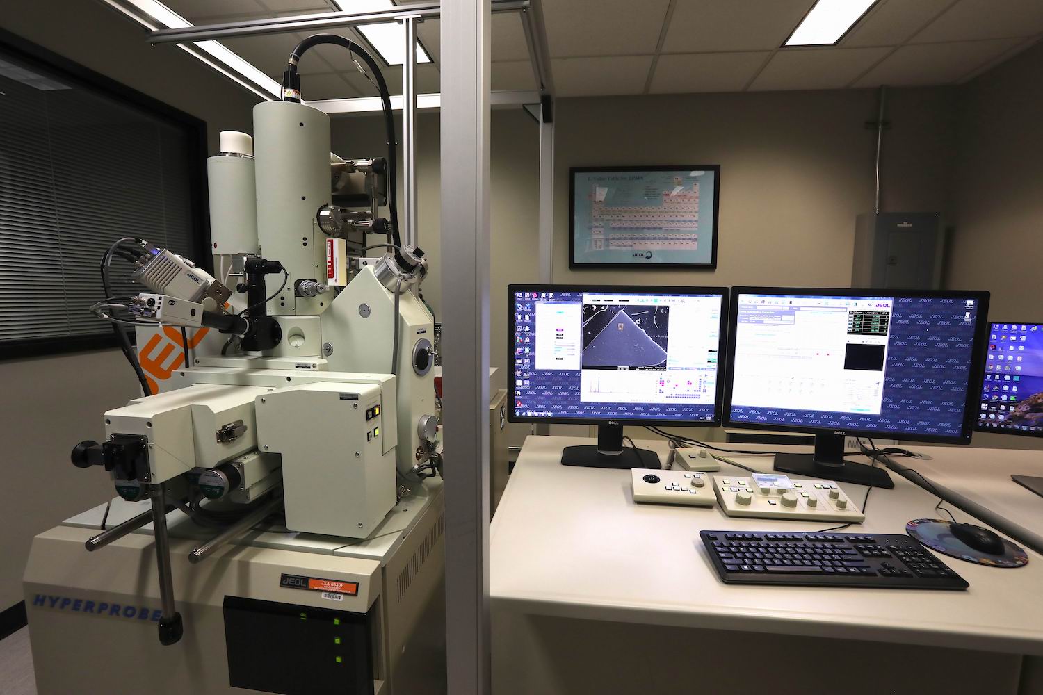
Purchased in December 2014, the JEOL JXA 8530F Hyperprobe was delivered in August 2015,was fully installed in December 2015 and it is managed by Dr. Gelu Costin (Gabi). The funding for the new equipment was entirely provided by Rice University.
The Jeol JXA 8530F Hyperprobe it is a fully-automated electron microprobe, customized for various EPMA applications. It has a digital imaging capability and can acquire digital backscattered images (in compositional and topographic modes), as well as secondary electron images. High-resolution digital X-ray intensity (element distribution) maps can be acquired using stage (for large maps) or beam (for small maps) scanning mode.
The Jeol JXA 8530F is equipped with a field emission gun (Schottky Field Emission Source), five wavelength dispersive spectrometers (WDS), and an integrated EDS detector. The spectrometer #1 has four analyzing crystals, while the spectrometers #2, #3, #4, and #5 have two analyzing crystals, each. Spectrometer #3 is an H-type, with a 100 mm radius of the Rowland circle, giving ~ three times higher count rates compared with an XCE spectrometer. The spectrometer #5 is an L-type, with large analyzing crystals (PETL, LiFL), suitable for trace element analysis, giving high X-ray intensities (without sacrificing the peak/background ratio), and higher spectral resolution. The customized configuration of the five spectrometers and the element range for each analyzing crystal are shown below:
|
Spectrometer |
Type |
Counter |
Analyzing Crystals |
|---|---|---|---|
| #1 | FCS | GPC | TAPJ, LDE2, PETJ, LDEB |
| #2 | XCE | XPC | LiF, PETJ |
| #3 | H | XM-86XPCH | PETHS, LiFHS |
| #4 | XCE | GPC | LDE1, TAPJ |
| #5 | L | XPC | PETL, LiFL |
|
Analyzing Crystal |
Element Range |
|---|---|
| LDE1 |
K⍺ lines of C, N, O, and F |
| LDE2 |
K⍺ lines of B, C, and N |
| LDEB |
K⍺ lines of Be and B |
| TAPJ |
K⍺ lines O-Si L⍺ lines Cr-Zr M⍺ lines La-Pt |
| PETJ, PETH, PETL |
K⍺ lines Si-Cr L⍺ lines Kr-Eu M⍺ lines Lu-Bi and Th-U |
| LiF, LiFH, LiFL |
K⍺ lines Ca-Rb L⍺ lines Sb-U |
X-ray counters: Spectrometers #1 and #4 use proportional gas flow counters. The gas used is P-10 (90% Argon – 10% Methane mixture) which is optimum for low energy X-ray detection. The spectrometers #2, #3, and #5 use sealed Xenon counters for the detection of higher energy X-ray photons.
EDS detector (integrated with the WDS system) – The EDS detector is a Silicon Drift X-ray Detector, with a 10mm² active area, 133eV resolution. Detection ranges from Boron through Uranium.
Cathodoluminescence (CL) detector (integrated with the WDS system)- XM-26740PCLI – Panchromatic Cathodoluminescence Image System provided by JEOL. The CL detector is integrated within the EPMA software and is able to acquire individual, high-resolution CL images (grayscale or fake-colors), and can be used synchronously with the WDS mapping.
NOTE! The element lists are set up in advance for most routine materials, and judgment based on experience is used to select a particular x-ray line and crystal for a given application. This is why the operator needs to know the application before judging the setup of the microprobe (beam current and size, standards to be used, X-ray lines, backgrounds, etc).
Other features of the JXA 8530F are:
- Integrated visible- reflected light microscopy at 400 x magnification
- Auto-focus system for optimal sample positioning
- Secondary electron detector
- Backscattered electron detector (annular solid-state type for compositional and topographical imaging)
- Magnification range 40X to 300,000X (144 steps)
- Specimen exchange for up to 100mm diameter x 50mm samples
- Computer automation operating through PC compatible JEOL software on a Windows Windows7 platform.
Our lab uses the Virtual WDS registered software (® Cambridge University) for simulation of X-ray peaks, backgrounds, interferences, and strategy of X-ray counting.
Accessory: JEE-420 vacuum evaporator: used for the carbon coating of samples.
After basic training and a first supervised EPMA session, the users are encouraged to work independently (unsupervised). Even during unsupervised sessions, the assistance is available, at any time during the workday. The users are encouraged to ask for assistance at any time they might need. For urgent assistance during the overtime EPMA sessions (overnight, weekends, and public holidays) please contact the lab manager at the contact number which will be provided.
General capabilities of our electron microprobe:
- Full quantitative analysis. All detectable elements (from Be to U) are quantified on a spot of ca.100 nm to 1μm diameter or larger. Detection limits range 30-100 ppm, depending on the element and settings.
- Rapid qualitative and /or semi-quantitative analysis, in EDS or WDS mode and phase identification
- Line analyses (rapid compositional profiles)
- High-resolution chemical mapping (in WDS or integrated WDS-EDS mode) of specimens on scales from hundred nm to ca 8 cm
- CL imaging
- Imaging specimens at micro-scale using backscattered electron (BSE) and secondary electron signal; Crystals below 1 micron in size (tens to hundreds of nanometers) are imaged with reasonable resolution
- Topographic imaging for unpolished samples
- Trace element analysis for particular elements in minerals (detection limits: 5-30 ppm, depending on the element, mineral, and settings of the counting time)
- Monazite age dating
EPMA Standards
Our current mineral standard collection includes natural and synthetic minerals, natural and synthetic glasses, and synthetic metals, from various sources:
- SPI Supplies (53 natural minerals and 44 synthetic metals)
- Agar Scientific (30 natural minerals)
- Smithsonian Institution (minerals, natural and synthetic glasses, synthetic REE phosphates)
- Rhodes University (mostly secondary mineral standards)
- University of Arizona (natural and synthetic minerals and glasses primary standards)
- In-house produced standards (e.g. Fe3N)
Our large collection of mineral and synthetic EPMA standards allows us to be committed to a wide area of EPMA applications.
Visit our EPMA standards here: Rice_EPMA_standards
Our EPMA standard collection (primary and secondary standards) is constantly growing, as more standards become available and get mounted.
Contact
Dr. Gelu (Gabi) Costin
EPMA Lab Manager
Department of Earth Science
Rice University
6100 Main Street, Keith-Wiess Geological Laboratory, MS-126
Houston, TX, 77005
e-mail: g.costin@rice.edu Phone: Office: 713 348 2054
